


Oral Surgery | Dental Implants - Charlotte, NC
Caring is Our Reputation
Over the span of 23 years, Sharma Oral Surgery has consistently thrived in transforming patients into friends and integral members of our extended family. Our approach is simple yet profound: we put your well-being above everything else. We take immense pride in nurturing enduring relationships with our patients, getting to know you beyond your surgical needs, and ensuring your comfort at every step of your treatment with us.
Driven by a commitment to providing predictable and top-notch solutions, we’re here to make every visit like a reunion with an old friend. Sharma Oral surgery isn’t just about surgical procedures; it’s about creating lasting connections with the people we are privileged to serve. When you step into our surgical office, you’ll immediately sense the warmth and genuine friendliness that has become our hallmark.
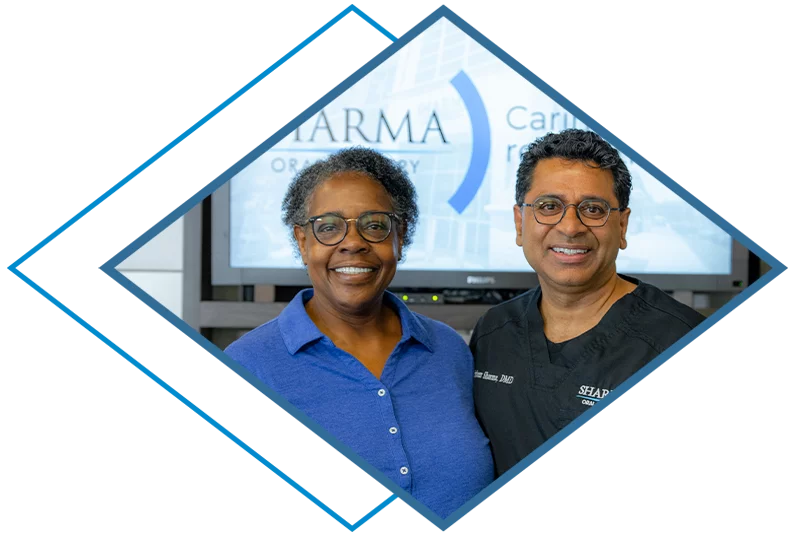
Meet Your Surgeon


Creating Healthier Futures,
One Procedure at a Time
View More Stories


Top-Rated Surgical Treatments

Full Mouth Dental Implants
Our full mouth dental implant solution is ideal for those with multiple missing teeth, providing a simple way to restore your complete smile. With our innovative Refresh Smile protocol, we can replace an entire arch of teeth using only 4-6 dental implants. This approach ensures precise placement, resulting in a brand-new, fully restored smile that looks and functions like natural teeth. Say goodbye to gaps and hello to a confident, fully functional smile with our Refresh Smile dental implant solution.

Single Tooth Implants
Single tooth implants are advanced tooth replacement solutions. A dental implant replaces the root of a tooth. This sturdy foundation supports a custom-designed crown, mimicking the appearance and function of a natural tooth. Single dental implants provide a long lasting and aesthetically pleasing solution for our patients with missing teeth, enhancing both oral health and the overall quality of the smile.

Wisdom Teeth Removal
Wisdom teeth, known as third molars, typically emerge in the late teens or early twenties. These molars can often cause discomfort and complications due to insufficient space in the jaw. We may recommend removing wisdom teeth to prevent infection, shifting and potential damage to neighboring teeth & nerves before they even erupt.

Oral Surgery
Oral surgery is a specialized field of dentistry that focuses on surgical procedures involving the mouth, teeth, jaws, and facial structures. It encompasses many procedures, from tooth extractions and dental implant placements to corrective jaw surgeries and treatment for oral diseases. Dr. Sharma is a highly trained board certified surgeon who addresses complex dental and facial issues, ensuring patients oral health and overall well-being.

Beyond Surgery
The Ole Study Club is an educational platform empowering doctors through engaging lectures and guest speakers, promoting lifelong learning and professional development principles. It creates a collaborative space for doctors to exchange ideas, stay updated on advancements, and enhance their practice with diverse topics and renowned speakers. As a valuable resource, it enables doctors to provide the highest quality care to their patients while staying at the forefront of their fields.